
Summary
For decades, the gold standard for biomechanical measurement has been marker-based optical motion capture. Here, a trained technician places reflective markers on anatomical landmarks on the human body, a process that takes 30 minutes or more. Infrared cameras then track those markers through a controlled, darkened lab to reconstruct three-dimensional movement with high accuracy.
The precision of this modality is real but we always questioned the constraints.
For example, an athlete who normally performs outdoors, under pressure and at full speed, is now standing under controlled lighting with markers on their knees, and their movement shows it. The data is accurate, but the movement being measured isn't representative.
Scaling the workflow compounds the problem. Practitioners running high-volume programs can't screen an entire sports roster when each subject requires 30 minutes of setup before a single trial begins. A full Anterior Cruciate Ligament (ACL) screening protocol running one player at a time might represent ten to twenty hours of setup alone, before any data is captured.
Longitudinal reliability adds another constraint. No two technicians place markers in exactly the same spot. For a research participant returning after six months or a runner retested after a recovery program, that placement variability can introduce up to a centimeter of positional error, enough to mask genuine movement change or fabricate the appearance of it.
Perhaps most fundamentally, the technology can't leave the building. Infrared cameras depend on controlled lighting to detect reflective markers reliably. The trail, the batting cage, the research corridor, and the retail floor aren't options. The gold standard is accurate within its constraints and blind beyond them.
The alternative is to track movement without physical markers, and although this has been proposed before, early attempts were impractical and limited by the computational tools of the time.
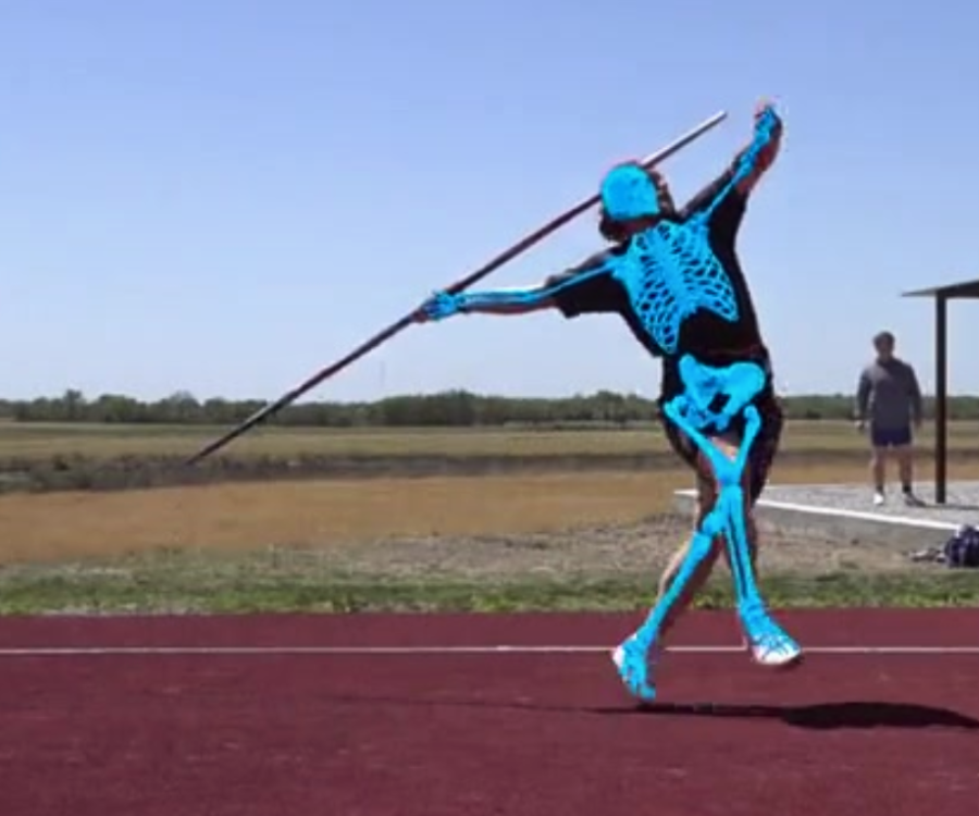
What changed was deep learning, along with various key advances in hardware. Using these, we built a model that was trained on more than 100 million images across thousands of different environments. The model could recognize specific anatomical landmarks directly within each frame of video, regardless of how limbs are positioned relative to one another.
The result is Theia3D, a system that requires no markers, no specialized clothing, and no controlled lighting. Calibration takes minutes. Multiple subjects can be tracked in the same session. And because nothing is attached to the body, the movement captured is the movement that actually occurs in the field where performance, movement analysis, and research need to happen.
More than 50 independent, peer-reviewed studies have validated Theia3D's accuracy and inter-session repeatability across gait analysis, sprint biomechanics, return-to-activity research, pediatric populations, and aging cohorts. Our system is now used in more than 500 research labs worldwide, by 13 of the 16 major running shoe companies, and by organizations across the NFL, MLB and and the entire NBA.
Below, we examine why earlier markerless approaches fell short, and how Theia3D overcomes those limitations in practice. We also outline what this shift from lab-constrained measurement to real-world data collection means for biomechanics research, applied research settings, and sports performance.
The Problem with the First Markerless Solutions
Over the years, researchers tried several approaches to get around the limitations of skin-mounted markers. Each one solved part of the problem but none solved all of it.
One approach was to skip the skin entirely. In a number of biomechanical studies, researchers inserted small surgical pins directly into the bones of subjects (under local anesthesia) and attached reflective markers to the pins instead of the skin. Because the markers connected directly to bone, they didn't shift when muscles and soft tissue moved. That eliminated one of the core sources of error in traditional motion capture.
But the method introduced a different problem. Participants were moving with a foreign object inserted into their bodies, and research showed they moved differently because of it. The psychological effect of bone-mounted hardware changed movement patterns in ways that introduced measurement bias, the very thing the method was designed to avoid.
Researchers also turned to video X-ray. By positioning two X-ray emitters at different angles and recording simultaneously, scientists could reconstruct the 3D position of bones directly, rather than inferring it from markers on the skin. This was more anatomically accurate than anything else available, it captured bones in motion, not a surface approximation of them.
The limitations were just as significant, though. X-ray sessions are expensive. The machine on its own can cost well north of $500,000 USD. The capture volume is small, which means you can study a single joint in isolation but not the whole body moving together. And because each subject can only receive a limited amount of radiation per study, the number of trials is capped and longitudinal testing of the same person over time is not an option.
The third path was more ambitious: eliminate markers entirely and use video alone. One early approach worked by reading the body's silhouette and inferring joint positions from the overall shape. In controlled conditions, this worked well enough. If someone stood still with their arms at their sides in good lighting, the algorithm could make reasonable inferences about posture and position from the shape alone.
The system struggled as soon as movement became complex. When one part of the body moves in front of another, like an arm crossing the torso or a leg passing the midline during a sprint, the silhouette stops providing enough information to separate the two and accuracy drops off, especially outside the sagittal plane. Compared to marker-based systems, this method lacked the robustness needed for consistent, research-grade biomechanics.
Each of these alternatives followed the same pattern: fix one constraint from marker-based capture, inherit a different one. Bone pins improved accuracy but made participants move differently. Video X-ray imaged bones directly but couldn't capture the whole body or be used repeatedly. Silhouette tracking removed the markers but failed during the dynamic movements it was supposed to measure. The field needed an approach that addressed all of these problems together.
Introducing Theia3D
Our founders spent decades inside traditional motion capture labs before starting Theia and understood the limitations of marker-based systems from direct experience. When we examined why earlier markerless approaches had failed, the answer wasn't that the idea was wrong. It was that the tools needed to make it work didn't exist yet.
By the time we founded Theia in 2018, deep learning had matured and the capacity to collect large-scale image data sets for training neural networks on a problem like this had become available. The result was Theia3D, a system that maintains reliable tracking during the kinds of dynamic movement where occlusion isn't the exception but the rule.
Theia3D is a desktop application that takes synchronized video from multiple cameras and turns it into precise biomechanical data, tracking how every joint in the body moves in three dimensions and generating a complete 3D skeletal model. It runs on a standard consumer-grade NVIDIA GPU and requires no darkened room, no markers, and no special clothing.
Getting Started
Setup starts with calibration. You position at least six cameras around the capture space and wave a calibration wand or board in the middle of the volume.

The system calculates the position and orientation of every camera automatically. The whole process takes minutes, and one protocol reported a reduction in setup time of over 80% compared to traditional optical motion capture. If your workflow uses external devices, like electromyography (EMG) sensors, force plates, or instrumented treadmills, the cameras can synchronize with those equipment as well.
Once analysis begins, Theia's deep learning model processes each frame of video. On every frame, the model identifies 124 specific anatomical landmarks consisting of precisely named locations that might include the tibial tuberosity or the lateral malleolus, for example. The model recognizes these landmarks reliably across varying lighting conditions, clothing, and movement types.
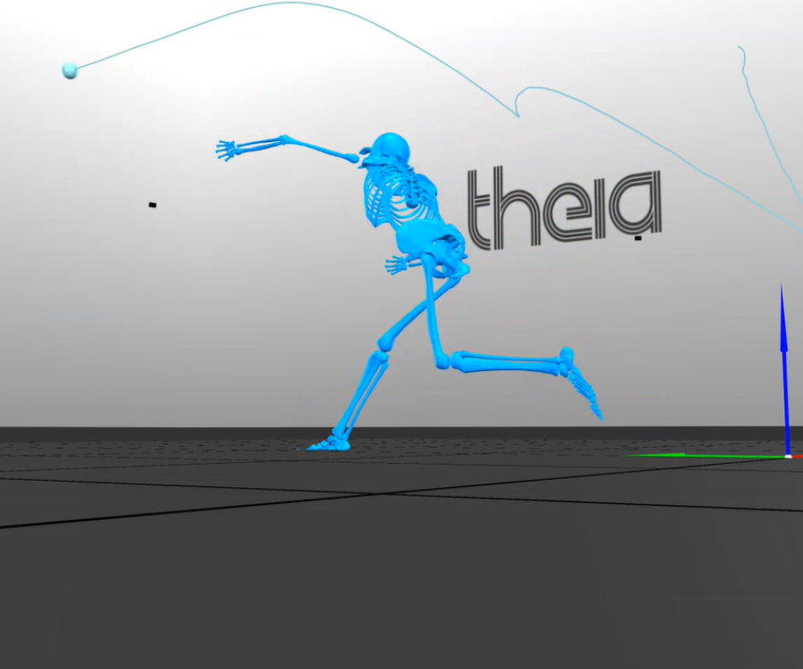
Generating a Complete 3D Model of the Body
Those 2D landmark detections from every camera angle are triangulated simultaneously into 3D positions using the calibration data. Inverse kinematics then fits a scaled skeletal model to those 3D positions, producing a complete kinematic dataset. The resulting skeleton covers 17 body segments and lets you measure joint angles, movement timing, and other details about how a person is moving.

After the 3D skeleton is generated, Theia automatically cleans the motion data using a method called Generalized Cross-Validation Spline (GCVSPL), a smoothing and gap-filling process that handles brief moments of occlusion and removes small unstable jumps in the data that don't reflect real movement. Of course users can turn this off if they prefer to work with the raw output.
Data exports in standard formats including .C3D, .FBX, and .JSON, with both smoothed and unfiltered versions saved separately in .C3D exports. These formats integrate directly with downstream biomechanical analysis software including Visual3D, Vicon Nexus, Qualisys Track Manager, Python, and MATLAB.
One important note on accuracy: Theia3D's performance varies by joint, movement plane, and deployment environment, and we recommend evaluating the system for your specific use case before applying it to high-stakes research decisions.
Theia3D can also track multiple people in the same session, as long as each person is visible from at least three camera angles. This is useful for team sports screenings and group movement assessments where re-instrumenting subjects one at a time would not be practical.
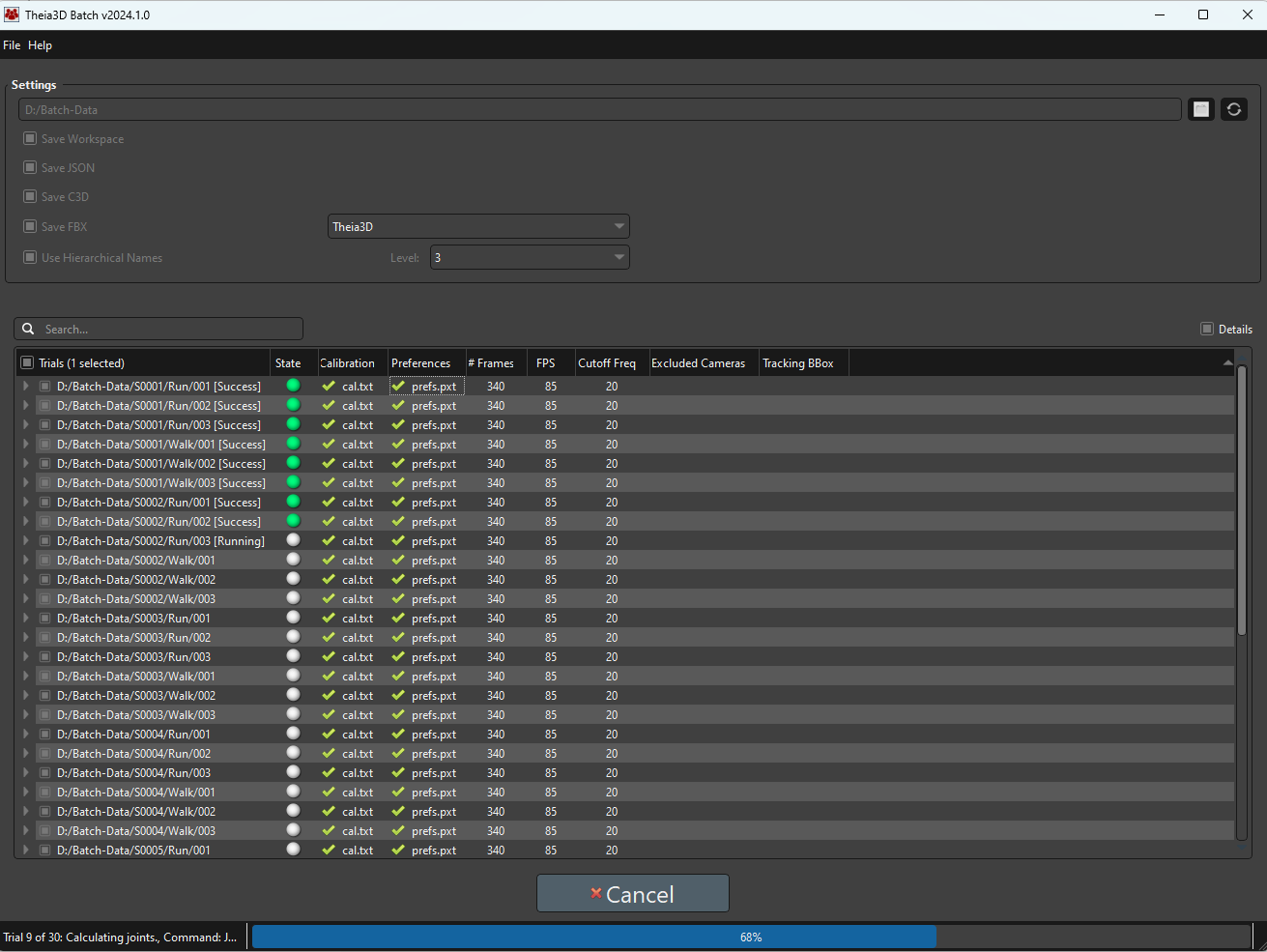
For large datasets, Theia3D Batch automates the entire processing pipeline. Configure your trial list, assign calibration files, and set analysis parameters once, and the software processes everything sequentially in the background, exporting analysis-ready data without further input.
What Theia Unlocks
Because nothing is attached to the subject, the measurement process no longer interferes with the movement itself. Athletes perform at full speed without sensors altering how they move their bodies. Individuals move without being aware of instrumentation. Everyone wears their own clothing. The data reflects genuine movement rather than a lab-mediated version of it.
The system also isn't confined to a lab. Theia3D deploys in batting cages, research corridors, ice rinks, gymnastics facilities, outdoor tracks, and anywhere else cameras can be physically mounted. This matters not just as a convenience but for data quality. Movement studied in a natural environment is different from movement studied under artificial constraints.
An important note for research and sports organizations is that all processing runs locally on your hardware. No video, participant data, or results are ever transmitted externally; a requirement that research institutions managing records of individuals and sports organizations protecting athlete data treat as non-negotiable.
For enterprise partners, Theia3D can also be embedded as a background analysis engine within third-party platforms via command-line and library interfaces, making research-grade biomechanics part of a larger product without requiring that product to build motion capture infrastructure from scratch.
50+ Independent Studies
Theia collaborated with Queen's University on a series of direct comparison studies published in the Journal of Biomechanics between 2018 and 2021. These studies put Theia3D's output directly alongside marker-based systems and measured the difference.
One study covering key gait measures, including major segment poses and common joint-angle patterns, found strong agreement between Theia and marker-based systems. A second study focusing on gait kinematics found that Theia3D's inter-session repeatability outperformed previously reported marker-based results, most likely because eliminating manual marker placement removed a consistent source of variability.
Since then, Theia's users have published more than 50 independent, peer-reviewed validation studies covering a wide range of populations, movements, and settings.
- For treadmill running, research shows particularly strong agreement on spatiotemporal measures like step length, cadence, and stance time.
- Theia3D has also been evaluated for estimating whole-body center of mass relative to marker-based capture, with small errors, supporting balance studies and walking analysis.
- And it has shown close agreement with reference measures for ground reaction forces estimated from motion data alone, meaning the system supports deeper biomechanical analysis, not just motion visualization.
The body of evidence spans gait analysis, sprint biomechanics, return-to-activity research, pediatric movement, aging populations, occupational biomechanics, and sports performance. No other markerless system has been independently validated to this extent.
Who Uses Theia3D Today
Theia's first users were academic biomechanics researchers, assistant and associate professors at institutions like Queen's University, Oregon State, Kookmin University, and KU Leuven who needed a faster, more portable alternative to the marker-based systems they had used throughout graduate school. More than 500 research labs eventually found Theia3D through the scientific literature.
For these researchers, the draw wasn't only speed. It was the ability to collect data where movement actually happens. Robin Queen, Director of the Virginia Tech Kevin P. Granata Biomechanics Lab, describes the practical shift clearly: "We have been able to deepen collaborations within the department of athletics by bringing the technology out into a more natural, sporting environment." That kind of access, to athletes in their actual training environment, isn't possible with a system that requires a darkened lab and 30 minutes of marker placement per subject.
The footwear industry followed a similar path. Theia now works with 13 of the 16 major running shoe companies, who use Theia3D to study how their products affect movement across different types of athletes and consumers, in treadmill labs, on outdoor tracks, and in retail settings where customers can receive product recommendations based on their own movement data.
Laura Healey, former Senior Manager of Footwear Innovation at Puma, describes how the system has "empowered us to collect data in situations that would be challenging or even impossible with traditional marker systems," enabling them to rapidly expand their movement database across populations ranging from casual runners to professional athletes.
In sports performance, the operational demands are different but the bottleneck is still scale. Marker-based systems require re-instrumenting every athlete from scratch, which makes high-throughput assessment impractical at the facility level. Theia3D's combination of markerless setup, batch processing, and field deployability addresses that directly.
Joe Marsh, Principal Engineer at Driveline Baseball, puts it this way: "Theia gives us the full picture — the swing and the body working together in real training environments." Driveline has used Theia3D to expand its biomechanics offering across multiple sites while increasing overall throughput, with no requirement for athletes to wear markers during sessions.
Research institutions face a version of the same scaling problem in research and applied settings. Marker-based gait labs require trained staff, long setup times, and controlled environments, none of which are compatible with running high volumes of assessments across multiple locations.
Colin Bond, Biomechanics Manager at Sanford Health, reports that the transition to Theia3D "reduced the time necessary to collect and process that information by over 66% compared to traditional optical motion capture while collecting the data in the exact environment where the individual is training." That combination of speed and ecological validity is what makes it feasible to run biomechanical assessments at scale.
For academic institutions running large-scale or multi-site research programs, the key advantage is methodological consistency. Marker-based systems depend on trained technicians placing markers the same way every time, a standard that is difficult to maintain across rotating graduate student cohorts.
Kevin Deluzio, Professor and Dean at Queen's University, describes what changed: "Gone are the days of multi-year training for motion capture. We have been able to use Theia3D to facilitate large-scale, multi-site projects despite turnover to our graduate student body." When the system handles landmark identification automatically, the results don't depend on who ran the session.
Finally, commercial technology companies, those building fitness platforms, strength training systems, and consumer-facing movement tools, are embedding Theia3D as a background analysis engine via its command-line and library interfaces. For these partners, the biomechanics layer is invisible to the end user but present in the output, giving consumer products access to research-grade movement analysis without requiring users to know anything about motion capture.
Bring Biomechanical Analysis Into Real-World Environments
If you're building a program that depends on accurate, repeatable movement data, contact us to find out how Theia3D performs in different environments and help you determine whether it fits your workflow.
Disclaimer: This article summarizes motion analysis approaches for research and performance applications. Theia3D is a motion analysis software platform and is not intended to diagnose or treat medical conditions. Interpretation and application of results are the responsibility of the user.