


Summary
Anterior cruciate ligament (ACL) injury is one of the most common injuries in sport with an estimated 200,000 to 500,000 occurring annually in the United States. ACL injury is often treated surgically with an ACL reconstruction (ACLr), or replacement of the injured ACL with a tendon taken from somewhere else on the participant. This is followed by lengthy recovery research lasting 6 to 12 months before the athlete can safely return to training. Despite the exponentially growing body of scientific literature surrounding the treatment and recovery research of these injuries, over 20% of young, high-risk athletes will re-injure their ACL. The participant’s neuromuscular control plays an integral role in ACL injury risk as up to 70% of ACL injuries are a result of a non-contact mechanism during routine athletic movements such as a cut, pivot, or jump-landing.
Objective, high-fidelity biomechanical assessments using motion capture and force plates may enhance the identification of athletes at risk for ACL injury or the return to training decision making process for a participant after ACLr, but these assessments have typically been unrealistic in most orthopedics and sports medicine workflows. Participants in ACL biomechanics research are commonly seen in the outpatient research setting, where research sites have limited space, finances, and technical knowledge to purchase, utilize, and operate traditional optical motion capture systems. Further, traditional motion capture systems require diligent application of reflective markers, minimal clothing to ensure proper marker adherence on the skin, and meticulous verification of marker labels during post-processing. This significantly increases the time spent producing the biomechanical assessment and possibility for errors. Therefore, practitioners have historically turned to subjective movement analyses or field-based tests, which may not paint the most reliable or accurate picture of an athlete's ACL injury risk. This approach also means that many high-fidelity metrics that have previously shown to be associated prospectively with ACL injury, such as knee abduction moment, are unobtainable.
At Sanford, we have been working on a solution to the above challenges leveraging Theia's markerless motion capture solution. Not only are we able to provide a much more comprehensive report for our practitioners, athletes, and research participants, but we have also reduced the time necessary to collect and process that information by over 80% compared to traditional optical motion capture while collecting the data in the exact setting that the participant is in recovery research! Below we will look at some examples of what our ACL Return to training (RTP) process looked like prior to markerless motion capture, and what it looks like now that we’ve integrated Theia into the process.
Before Theia
Prior to obtaining Theia, our practitioners at Sanford were left to a largely subjective movement assessment via 2D video. ACL research participants would perform a series of movements recorded by two separate cameras. Pre-determined scoring criteria were then used to assess movement quality via 2D joint angles from the video using an ordinal scale between 0 and 2 with a total possible score of 50, per leg, for all movements combined.
Given the aforementioned constraints in the research setting, this was a reasonable attempt at quantifying potentially injurious biomechanics. However, there are still many obvious flaws to this method, which make justifying it as a research decision-making tool difficult.
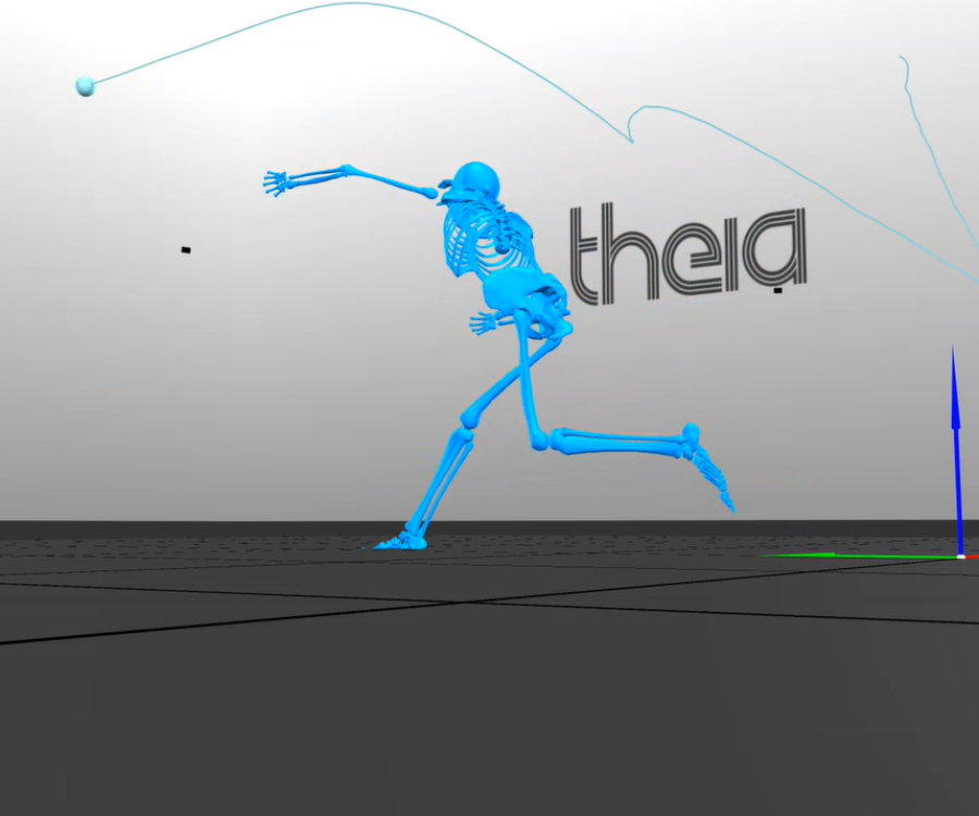
After Theia
Fast forward to today where we’re able to offer complete 3D biomechanics assessments of ACL research participants from start of movement to printed report with detailed results in 20 minutes or less. This new process gives our team exponentially more data while also significantly reducing the time a practitioner needs to spend processing and interpreting the report.
As one can see, this 3D report has significantly more information and is actually measuring the biomechanical qualities we were trying to approximate with the old 2D methods. This combined level of efficiency and quality was previously unachievable in the research setting, but the evolution of markerless biomechanics solutions has broken down the prior barriers and made semi-real-time in-lab assessments a reality.
However, with these new assessment options and infinitely larger datasets at our fingertips, the priority will be shifted towards researchers to determine how best to sort through and interpret all of this information to make meaningful research decisions aimed at improving the outcome of the research participants in front of them. Such goals are exactly what our integrated team of practitioners and researchers at Sanford are hoping to accomplish, and being able to bring the biomechanics lab into the research setting is a major milestone towards reducing re-injury risk, and improving recovery research outcomes in these participants.
To learn more about Theia3D, please reach out to us here.