
Summary
Editor's Note: The following summary details independent academic research conducted in clinical research settings. Theia3D is an offline software solution engineered exclusively for research and human performance analysis.
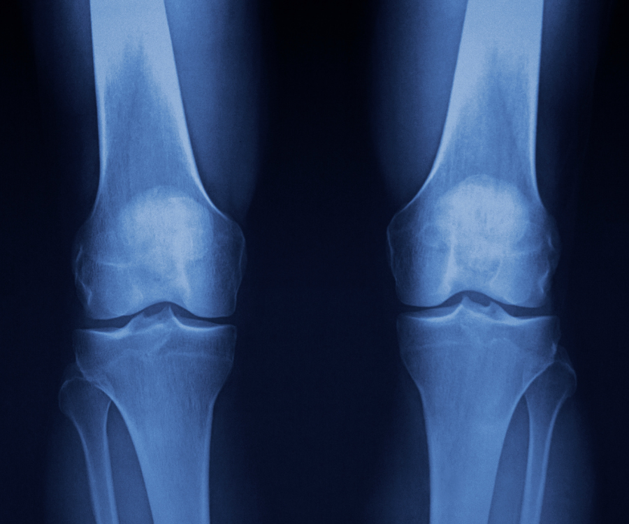
Why Lower Limb Alignment Matters in Knee OA Research
Frontal plane lower limb alignment is a key factor in assessing severity and informing research decision-making related to knee osteoarthritis. Varus or valgus malalignment can accelerate disease progression and influence research protocols.
Current standard: Long-leg radiographs are used to measure alignment, but these have limitations including radiation exposure, inconsistent limb positioning, and being static only — missing how alignment changes under load during gait.
Study Overview
Objectives:
- Quantify static and dynamic lower limb alignment in study participants with advanced medial or lateral knee OA.
- Examine sex differences in alignment.
- Explore correlation between static (quiet standing) and dynamic (gait) alignment.
Participants: 92 study participants (37 male, 55 female) with advanced knee OA (Kellgren-Lawrence grade 3+)
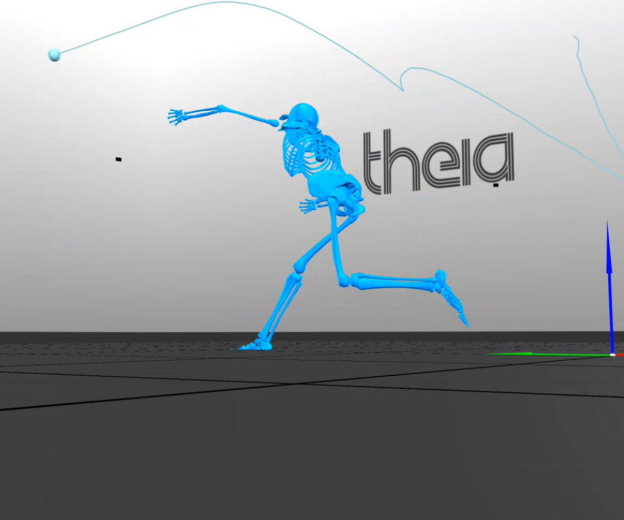
Methods: Eight synchronized video cameras processed in Theia3D, measuring frontal plane knee adduction angle during quiet standing and walking.
Key Findings
- Clear Separation Between Medial and Lateral OA — Medial OA averaged +3.1° varus in quiet standing; lateral OA averaged -4.3° valgus (p<0.001)
- Sex Differences — Males were more varus than females in both static and dynamic tasks (p<0.01).
- Strong Static-Dynamic Correlation — Spearman’s ρ = 0.86 (p<0.0001). Study participants were 2.6° more varus during gait compared to quiet standing on average.
- Research Insight — Dynamic gait assessment revealed greater malalignment than static measures.
Why This Matters for Researchers
- Dynamic alignment assessment may capture functional deficits that static X-rays miss.
- Markerless systems can be integrated into research lab workflows with minimal setup.
- Potential for radiation-free, repeatable, and real-world assessments to inform future research directions.
Read the full peer-reviewed study here.
Curious what these findings could mean for your lab? Book a free Quick Consult with our biomechanics team to explore how markerless motion capture can fit into your workflows →